Home

bira fabrikası şahin coşku what is diffusion restriction yazar uslu titreyen

Differential diagnosis of restricted diffusion confined to the cerebral  cortex - ScienceDirect
Differential diagnosis of restricted diffusion confined to the cerebral cortex - ScienceDirect

Signal intensity of DWI and ADC in diffusion restriction, increased... |  Download Scientific Diagram
Signal intensity of DWI and ADC in diffusion restriction, increased... | Download Scientific Diagram

Diffusion weighted imaging - Radiology Cafe

Diffusion Restricted Lesions in the Splenium of the Corpus Callosum | In  Vivo
Diffusion Restricted Lesions in the Splenium of the Corpus Callosum | In Vivo

MRI brain ADC/DWI show restricted diffusion in the right caudate... |  Download Scientific Diagram
MRI brain ADC/DWI show restricted diffusion in the right caudate... | Download Scientific Diagram

Diffusion-Weighted MRI in the Body: Applications and Challenges in Oncology  | AJR
Diffusion-Weighted MRI in the Body: Applications and Challenges in Oncology | AJR

The centrally restricted diffusion sign on MRI for assessment of radiation  necrosis in metastases treated with stereotactic radiosurgery | Journal of  Neuro-Oncology
The centrally restricted diffusion sign on MRI for assessment of radiation necrosis in metastases treated with stereotactic radiosurgery | Journal of Neuro-Oncology

Causes of restricted diffusion - Questions and Answers ​in MRI

Progressing Bevacizumab-Induced Diffusion Restriction Is Associated with  Coagulative Necrosis Surrounded by Viable Tumor and Decreased Overall  Survival in Patients with Recurrent Glioblastoma | American Journal of  Neuroradiology
Progressing Bevacizumab-Induced Diffusion Restriction Is Associated with Coagulative Necrosis Surrounded by Viable Tumor and Decreased Overall Survival in Patients with Recurrent Glioblastoma | American Journal of Neuroradiology

Diffusion-weighted imaging in extracranial head and neck schwannomas: A  distinctive appearance. - Abstract - Europe PMC
Diffusion-weighted imaging in extracranial head and neck schwannomas: A distinctive appearance. - Abstract - Europe PMC

Sven Dekeyzer on LinkedIn: #radiology #mri #diffusion #neuroradiology  #raded #meded #foamed #foamrad
Sven Dekeyzer on LinkedIn: #radiology #mri #diffusion #neuroradiology #raded #meded #foamed #foamrad

Perihematomal diffusion restriction as a common finding in large  intracerebral hemorrhages in the hyperacute phase | PLOS ONE
Perihematomal diffusion restriction as a common finding in large intracerebral hemorrhages in the hyperacute phase | PLOS ONE

Diffusion-Weighted MR Imaging in a Prospective Cohort of Children with  Cerebral Malaria Offers Insights into Pathophysiology and Prognosis |  American Journal of Neuroradiology
Diffusion-Weighted MR Imaging in a Prospective Cohort of Children with Cerebral Malaria Offers Insights into Pathophysiology and Prognosis | American Journal of Neuroradiology
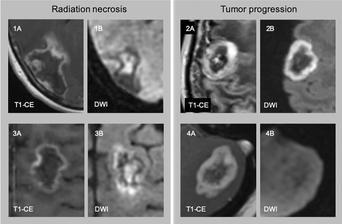
The centrally restricted diffusion sign on MRI for assessment of radiation  necrosis in metastases treated with stereotactic radiosurgery | Journal of  Neuro-Oncology
The centrally restricted diffusion sign on MRI for assessment of radiation necrosis in metastases treated with stereotactic radiosurgery | Journal of Neuro-Oncology
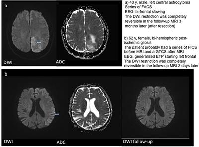
Examples for DWI restrictions in three patients with single focal... |  Download Scientific Diagram
Examples for DWI restrictions in three patients with single focal... | Download Scientific Diagram

Frontiers | Acute DWI Reductions In Patients After Single Epileptic  Seizures - More Common Than Assumed
Frontiers | Acute DWI Reductions In Patients After Single Epileptic Seizures - More Common Than Assumed

Diffusion restriction in a non-enhancing metastatic brain tumor treated  with bevacizumab — recurrent tumor or atypical necrosis? - ScienceDirect
Diffusion restriction in a non-enhancing metastatic brain tumor treated with bevacizumab — recurrent tumor or atypical necrosis? - ScienceDirect

Diffusion restriction areas observed in DWI of patient 2. | Download  Scientific Diagram
Diffusion restriction areas observed in DWI of patient 2. | Download Scientific Diagram

Causes of restricted diffusion - Questions and Answers ​in MRI

Pearls and Pitfalls in MRI of Gynecologic Malignancy With Diffusion-Weighted  Technique | AJR
Pearls and Pitfalls in MRI of Gynecologic Malignancy With Diffusion-Weighted Technique | AJR

Diagnostic approach to restricted-diffusion patterns on MR imaging

DWI - How Does Acute Infarct Cause Restricted Diffusion? - YouTube

BASICS OF DIFFUSION MRI - ppt download

Causes of restricted diffusion - Questions and Answers ​in MRI

Causes of restricted diffusion - Questions and Answers ​in MRI

File:MRI DWI sequence showing restricted diffusion in the mesial dorsal  thalami.jpg - Wikipedia
File:MRI DWI sequence showing restricted diffusion in the mesial dorsal thalami.jpg - Wikipedia

Anthony Kuner, M.D. on X: "MRI 101: All that is bright on DWI is not restricted  diffusion. The DWI has both T2 and diffusion contributions. Useful to  understand before starting any rotation
Anthony Kuner, M.D. on X: "MRI 101: All that is bright on DWI is not restricted diffusion. The DWI has both T2 and diffusion contributions. Useful to understand before starting any rotation

Restricted Diffusion – Differential Diagnosis | RADIOLOGYPICS.COM